Our first integrated diagnosis platform using in vivo pathology imaging diagnostic technology based on ultrahigh-resolution OCT (HROCT) and ultrahigh-depth OCT (HDOCT) can be extended to multiple clinical fields. Our ultra-compact probe of 130-230μm (suitable for different kinds of puncture needles) and 1μm pathological-level resolution can better analyze lesions compared to traditional OCT.
The platform can accurately guide various thermal ablation treatments, ensuring 5mm+ minimal resection margin to eliminate tumor residues and block local tumor progression and significantly remove the limitation that MRI has with only 1mm resolution. The market of neurosurgical laser interstitial thermal therapy (LITT, also called MRI-guided laser ablation) — a minimally invasive surgical technique that helps surgeons precisely target and ablate tumor tissue — is still blank. Our HROCT platform allows precise 1μm, real-time guided laser ablation. The first-generation principle prototype has been completed.
Neurosurgery, worldwide clinical pitfalls of LITT
Pain points summary, from 15 years of global LITT clinical experience:
Successful treatment of LITT depends on accurate placement of the LITT probe and complete ablation of the entire cancer volume:
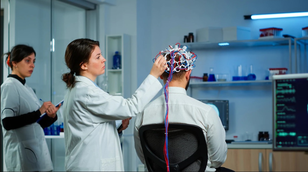
Ablation efficacy evaluation of LITT: During laser ablation, besides visualizing the treatment volume via MRI, the current practice provides very limited in situ real-time feedback on whether the entire lesion is ablated intact, other than temperature mapping. So far, there is no 1μm high-resolution technology which can assess in real-time whether there is residual lesion to guide further ablation, and whether the probe is close to critical vessels which should not be ablated.
Accurate placement of LITT probe: The LITT probe is placed in the operating room, but its accurate position cannot be confirmed until an intraoperative MRI scan is performed. If inaccurate, the probe should be repositioned and MRI should be rescanned, which is not only time-consuming and expensive but also increases the risk of potential injury to patient, especially for fragile and sensitive deep brain structures.
Principle prototype ready
Our platform allows precise 1μm and real-time guided laser ablation and is suitable for multiple organs. At present, our smart needle probe (SNP) technology can only realize the need for pathological diagnosis at any time with the multi-degree of the freedom movement. If combined with LITT products, we also hope to develop robots that can integrate into the catheter of LITT while pathological diagnosis, realizing the theranostic clinical purpose. The first-generation principle prototype has already been completed.
Our fiber-optic robot can be used in complex and restricted human diagnosis working environments, which cannot be handled by existing commercialized continuum robots. It has an ultra-fine mechanical body with a 600μm outer diameter and a 360° wide-angle freedom of movement and is controlled by special NdFeB programmed magnetic polarities, which is currently the world’s top soft robot.